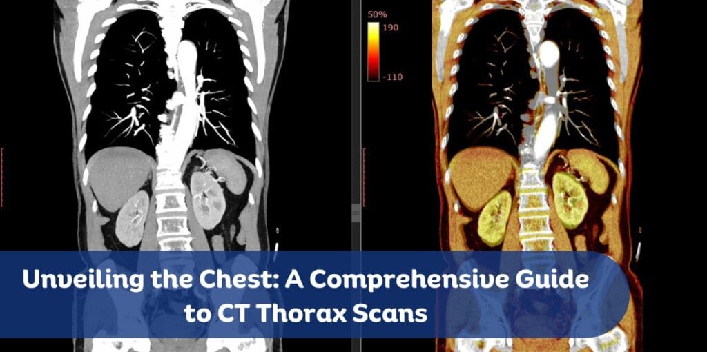
Unveiling the Chest: A Comprehensive Guide to CT Thorax Scans

A CT thorax scan, also known as a chest CT scan, is a powerful diagnostic tool that provides detailed images of the chest, including the lungs, heart, blood vessels, and bones. This advanced imaging technique is crucial for diagnosing and monitoring various conditions, offering a clearer picture than standard X-rays. In this blog post, we will explore the importance of CT thorax scans, the procedure, preparation, and what to expect during and after the scan.
Understanding CT Thorax Scans
A CT (computed tomography) thorax scan uses X-rays and computer technology to create cross-sectional images of the chest. These images provide detailed information about the structures within the chest, allowing healthcare providers to diagnose and monitor a wide range of conditions. The scan can detect abnormalities that may not be visible on a standard chest X-ray, making it an invaluable tool in modern medicine.
Why You Might Need a CT Thorax Scan
There are several reasons why a healthcare provider might recommend a CT thorax scan:
- Detecting Infections: CT scans can identify lung infections such as pneumonia, tuberculosis, and fungal infections. They provide detailed images that help in assessing the extent and severity of the infection.
- Evaluating Injuries: After trauma to the chest, a CT scan can reveal injuries to the lungs, ribs, heart, and blood vessels. This is crucial for determining the appropriate treatment plan.
- Diagnosing Diseases: CT scans are essential for diagnosing lung cancer, pulmonary embolism, and other lung diseases. They can detect tumors, blood clots, and other abnormalities with high precision.
- Monitoring Conditions: For chronic conditions like pulmonary fibrosis, chronic obstructive pulmonary disease (COPD), and interstitial lung disease, CT scans help track disease progression and response to treatment.
- Guiding Procedures: CT scans assist in guiding biopsies and other interventional procedures by providing precise images of the target area.
Preparing for the Scan
Preparation for a CT thorax scan is generally straightforward, but it may vary depending on whether a contrast dye will be used. Here are some common preparation steps:
- Dietary Restrictions: If a contrast dye is used, you may be asked to avoid eating or drinking for a few hours before the scan. This helps reduce the risk of nausea and ensures clear images.
- Medications: Continue taking your regular medications unless instructed otherwise by your healthcare provider. Inform your provider about any allergies, especially to iodine or contrast dye.
- Clothing and Accessories: Wear comfortable clothing and remove any metal objects, such as jewelry, eyeglasses, and hairpins, as they can interfere with the imaging process.
During the Scan
The CT thorax scan procedure is quick and painless. Here’s what you can expect:
- Arrival: Upon arrival at the imaging center, you will check in and provide any necessary medical history. You may be asked to sign a consent form.
- Preparation: You may need to change into a hospital gown and remove any metal objects. If a contrast dye is used, it may be administered orally or through an intravenous (IV) line.
- Positioning: You will lie on a motorized table that slides into the CT scanner, a large machine with a tunnel in the center. The technologist will position you correctly and may use straps or pillows to help you stay still.
- Scanning: The technologist will operate the scanner from a separate room but will be able to see, hear, and speak with you throughout the procedure. You may be asked to hold your breath for short periods to ensure clear images. The scan itself usually takes just a few minutes, although the entire process may take about 30 minutes.
After the Scan
Once the scan is complete, you can typically resume your normal activities. If a contrast dye was used, you might be advised to drink plenty of fluids to help flush it out of your system. Your healthcare provider will review the images and discuss the results with you, which can help guide further treatment or diagnostic steps.
Understanding the Results
The images from a CT thorax scan are interpreted by a radiologist, a doctor specialized in reading and analyzing medical images. The radiologist will send a report to your healthcare provider, who will discuss the findings with you. The results can reveal a variety of conditions, including:
- Lung Diseases: Such as pneumonia, tuberculosis, lung cancer, and COPD.
- Heart Conditions: Including heart enlargement, pericardial effusion, and congenital heart defects.
- Blood Vessel Abnormalities: Such as pulmonary embolism, aortic aneurysm, and vascular malformations.
- Bone and Soft Tissue Issues: Including rib fractures, spinal abnormalities, and soft tissue masses.
Risks and Considerations
While CT thorax scans are generally safe, there are some risks and considerations to keep in mind:
- Radiation Exposure: CT scans involve exposure to a small amount of radiation. While the risk is minimal, it is important to discuss any concerns with your healthcare provider, especially if you are pregnant or have had multiple scans.
- Allergic Reactions: Some people may have allergic reactions to the contrast dye used in CT scans. Inform your healthcare provider if you have a history of allergies to iodine or contrast materials.
- Kidney Function: The contrast dye can affect kidney function, particularly in individuals with pre-existing kidney conditions. Your healthcare provider may perform blood tests to check your kidney function before administering the dye.
Advances in CT Technology
Advancements in CT technology have significantly improved the quality and safety of thorax scans. Modern CT scanners use lower doses of radiation while providing high-resolution images. Additionally, new techniques such as dual-energy CT and 4D CT offer enhanced imaging capabilities, allowing for better diagnosis and treatment planning.
Conclusion
A CT thorax scan is a vital diagnostic tool that provides detailed images of the chest, helping healthcare providers diagnose and monitor a wide range of conditions. Understanding the procedure, preparation, and what to expect can help alleviate any concerns and ensure a smooth experience. With ongoing advancements in CT technology, these scans continue to play a crucial role in modern medicine, offering hope and clarity to patients and healthcare providers alike.
